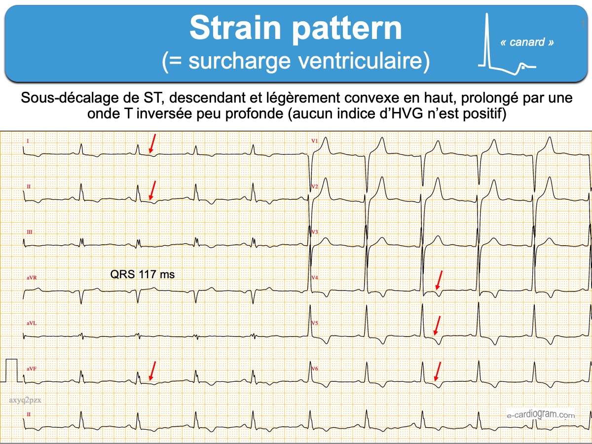
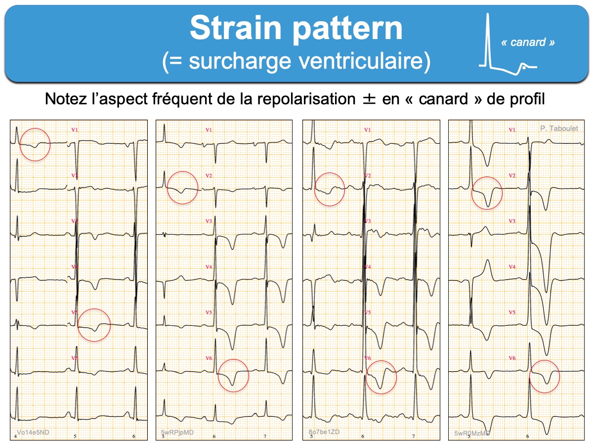
Terme anglais (strain : « contrainte ») qui correspond à un aspect type (pattern) de la repolarisation évocateur d’une surcharge ventriculaire. Il s’agit d’une anomalie secondaire de la repolarisation qui accompagne une hypertrophie/dilatation ventriculaire gauche ou droite [1].
L’ECG présente :
- un/des indice(s) d’hypertrophie ventriculaire positif(s) (mais pas toujours)
- un sous-décalage de ST en dérivations où l’onde R est proéminente, modéré, à partir d’un point J généralement peu abaissé et suivi, par une onde T inversée soit exclusivement négative, soit négative mais diphasique et qui se redresse rapidement dans sa partie terminale pour retourner à la ligne de base ou devenir brièvement positive (« overshoot ») [2]. La repolarisation ressemble à un canard vu de profil (ou à un cygne si le strain est très prononcé).
- L’intervalle QT est normal (≠ hypokaliémie ou amiodarone).
Sa présence témoigne d’un “remodelage électrique du cœur”. Il aggrave le pronostic des patients (ex. HTA [3], valvulopathies aortiques [4]).
Voir Surcharge ventriculaire gauche ou Surcharge ventriculaire droite
[1] Ehara S, Hasegawa T, Matsumoto K, et al. The strain pattern, and not Sokolow-Lyon electrocardiographic voltage criteria, is independently associated with anatomic left ventricular hypertrophy. Heart Vessels. 2014 Sep;29(5):638-44.
[2]Wang K et al. ST-segment elevation in conditions other than acute myocardial infarction. N Engl J Med 2003; 349(22): 2128-35. (téléchargeable)
Salles G, Cardoso C, Nogueira AR, Bloch K, Muxfeldt E. Importance of the electrocardiographic strain pattern in patients with resistant hypertension. Hypertension. 2006 Sep;48(3):437-42. (téléchargeable)
[3] Rautaharju PM, Soliman EZ. Electrocardiographic left ventricular hypertrophy and the risk of adverse cardiovascular events: a critical appraisal. J Electrocardiol. 2014 Sep-Oct;47(5):649-54.